



Explore the Tumor Microenvironment
Recreate key cellular and extracellular interactions
that shape tumor behavior and treatment response.
Build the Tumor Microenvironment
On-Chip
The TME-Chip builds the tumor microenvironment directly on the chip. Tumor, stromal, and immune cells can be co-cultured and self-assemble into 3D spheroids within a tunable extracellular matrix (ECM), so cell-cell and cell-matrix interactions, and their effects on tumor behavior and treatment response, can be studied in one model with live imaging and on-chip immunostaining.
On-Chip Spheroid Formation and Long-Term Culture
Tumor models assemble in place and can be followed over time.
Tumor cells, alone or with fibroblasts or other stromal cells, are loaded as a single cell suspension and self-assemble into spheroids in situ, with no separate pre-formation or transfer steps. Under perfusion, cultures can be maintained for up to 2 weeks, long enough to follow spheroid growth, matrix contraction, and stromal activation in the same chamber by live imaging.

BxPC-3 tumor cells and pancreatic stellate cells assemble into tumor spheroids in situ on the TME-Chip, with no pre-forming or transfer steps. The same chamber is shown at Days 4, 6, 9, and 11 of perfused culture, as the constructs enlarge and the surrounding matrix contracts. Scale bar, 100 µm.
Tumor–Immune Interactions
and Immune Evasion
Observe how immune cells infiltrate, engage, and are restrained by tumor tissue.
The TME-Chip supports the study of immune cell migration through the matrix toward tumor cells. Immune cells loaded via the side channel migrate through the matrix toward the tumor compartment. Live imaging can be used to follow immune cell migration, proximity to tumor cells, infiltration, and tumor cell viability over time. Comparing conditions with and without stromal cells or immunomodulatory agents helps separate tumor-intrinsic resistance from microenvironment-driven immune suppression. Varying ECM composition or density show how the matrix affects immune cell entry.
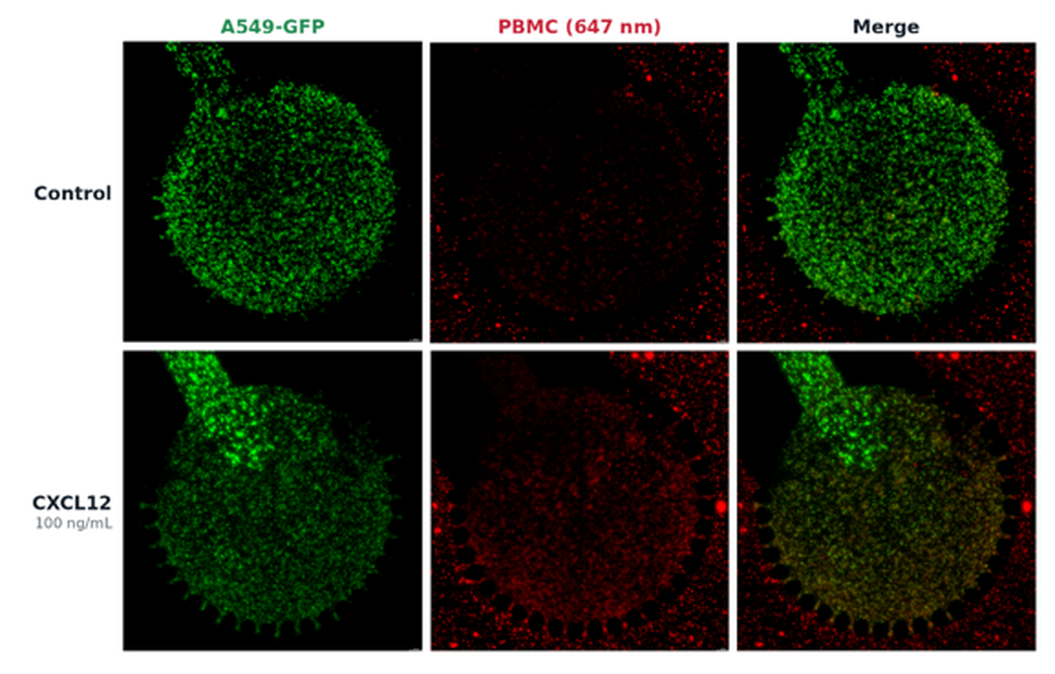
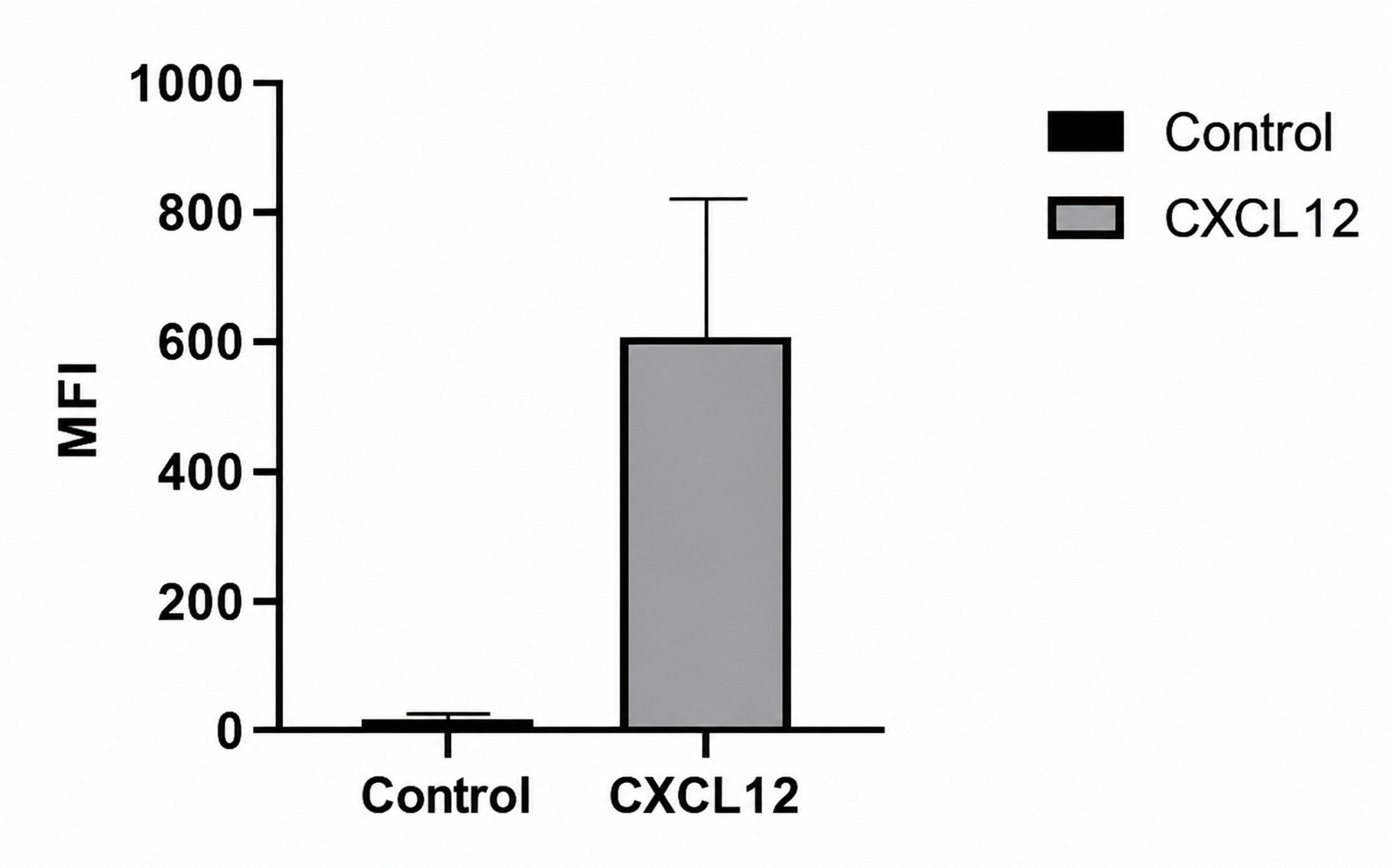
Chemokine-driven infiltration of immune cells into 3D tumor tissue on the TME-Chip.
A549-GFP lung cancer cells (green) were cultured in Matrigel in the tumor compartment, with or without CXCL12, as a chemoattractant. Primary human peripheral blood mononuclear cells (PBMCs), pre-labeled with a far-red cytoplasmic tracer (red), were introduced through the side channel. After 5 days, PBMC fluorescence intensity within the tumor compartment with CXCL12 increased relative to the untreated control, which remained near background.


A549 lung cancer cells (green) were cultured on the TME-Chip with IL-1β, and Jurkat T cells (red) were then added to the side channel. After 48 h, Jurkat T cells were seen clustered around A549 cells.
Cancer-Associated Fibroblasts and Extracellular Matrix
Examine how fibroblasts and a tunable ECM shape tumor behavior.
Cancer-associated fibroblasts regulate tumor growth, immune activity, and drug penetration through cell–cell contact, secreted factors, and ECM remodeling. On the TME-Chip, ECM composition can also be modulated independently by varying matrix concentrations or incorporating collagen or other matrix proteins. Comparing tumor phenotypes and immune cell infiltration across cell and matrix conditions helps to distinguish the effects of fibroblasts from those of the ECM itself.
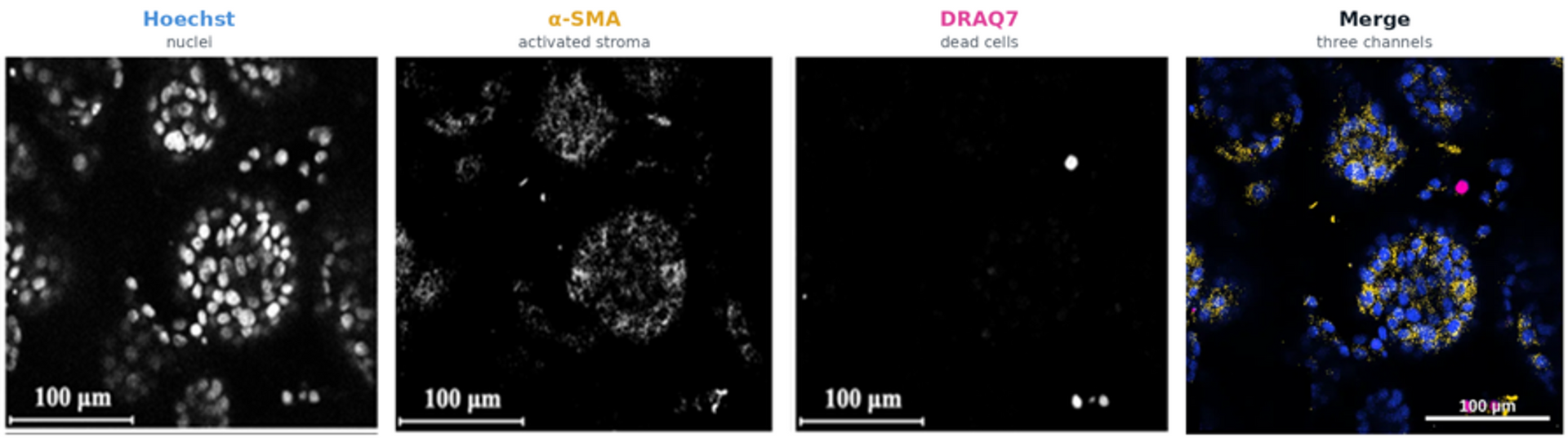
Activated stroma in a pancreatic cancer–stellate cell co-culture on the TME-Chip.
Human BxPC-3 pancreatic adenocarcinoma cells and pancreatic stellate cells (PSCs) were co-cultured under continuous perfusion for 11 days. On Day 11, α-SMA-positive PSCs were found intermixed with tumor cell clusters, indicating stromal activation. Few DRAQ7-positive cells were seen, suggesting most cells remained viable. Scale bar, 100 µm.
Contact Us
Connect with Pythia Biotech to learn more about our products and services,
explore partnering opportunities, or discuss potential collaborations.